Surgery removes fragments, corrects incongruity, and derides damaged cartilage, but it is rehabilitation that determines whether a dog returns to comfortable function or develops chronic compensatory dysfunction. Among dogs undergoing arthroscopic surgery for elbow dysplasia, those receiving structured post-operative rehabilitation demonstrate measurably better outcomes than those managed with rest-and-gradual-return protocols alone. For the substantial population of dogs managed conservatively, rehabilitation provides the primary means of maintaining muscle mass, joint mobility, and functional comfort across years of progressive osteoarthritis. Despite this, formal rehabilitation remains underutilized in veterinary orthopedics, partly because evidence has been slow to accumulate and partly because access to qualified canine rehabilitation practitioners remains uneven.
Why Rehabilitation Matters for Elbow Dysplasia
The canine elbow bears roughly 60% of body weight during standing and considerably more during movement, particularly during deceleration, turning, and downhill locomotion. Elbow dysplasia disrupts normal joint mechanics, creating a cascade of compensatory changes that extend well beyond the affected joint.
The Compensation Cascade
A dog with unilateral elbow pain instinctively shifts weight to the contralateral forelimb and hindquarters. Within weeks, this weight redistribution produces measurable changes:
- Forelimb muscle asymmetry: The affected limb loses muscle mass (particularly triceps and extensor groups) while the compensating limb develops relative hypertrophy.
- Contralateral overload: The sound forelimb absorbs increased forces, accelerating any subclinical pathology in that joint. Bilateral ED, present in 50-60% of affected dogs, makes this particularly problematic.
- Hindquarter shifting: Dogs redistribute weight posteriorly, increasing loading on hip and stifle joints. In breeds prone to concurrent hip dysplasia, this compensation accelerates hip disease progression.
- Cervical and thoracic spine strain: Altered head carriage and forelimb gait patterns increase strain on cervical and thoracic spinal musculature, sometimes producing secondary pain sources.
- Core weakness: Reduced overall activity levels lead to progressive core muscle deconditioning, further destabilizing the entire musculoskeletal system.
Rehabilitation addresses this entire cascade, not merely the primary joint. A program focused exclusively on the elbow while ignoring systemic compensation fails to restore functional movement patterns and leaves the dog vulnerable to secondary problems.
Research Insight: Post-Surgical Rehabilitation Outcomes
Muller et al. (2019) compared outcomes in dogs following arthroscopic treatment of medial coronoid disease. Dogs receiving structured rehabilitation (hydrotherapy, therapeutic exercises, manual therapy) showed significantly improved peak vertical force at 12 weeks compared to dogs managed with activity restriction alone. The rehabilitation group achieved 92% of normal limb loading versus 78% in the control group. Similar findings have been reported for post-osteotomy rehabilitation, though sample sizes remain small.
Post-Surgical Rehabilitation Protocols
Rehabilitation after elbow dysplasia surgery follows a phased approach, progressing from tissue protection through active recovery to return to function. The specific protocol depends on the surgical procedure performed, but the general framework applies across arthroscopic fragment removal, osteotomy procedures, and UAP fixation.
Phase 1: Protection and Pain Control (Days 1-14)
The immediate post-operative period focuses on tissue healing, pain management, and preventing muscle atrophy without stressing surgical repairs. Activity is restricted to leash walks for elimination only (5-10 minutes, 3-4 times daily). Cryotherapy (ice packs wrapped in a towel, 15-20 minutes, 3-4 times daily) reduces inflammation and provides analgesia. Gentle passive range of motion (PROM) exercises begin at 3-5 days post-surgery, within the comfortable range only. No resistance, no weight-bearing exercises.
Phase 2: Early Active Recovery (Weeks 2-6)
Controlled leash walks increase gradually (15-20 minutes, 2-3 times daily by week 4). Introduce underwater treadmill sessions at low speed and reduced water level, allowing partial weight bearing with buoyancy support. Active range of motion exercises replace passive techniques. Begin gentle weight-shifting exercises on stable surfaces. Heat therapy before exercise sessions improves tissue extensibility. Continue cryotherapy after activity.
Phase 3: Strengthening and Proprioception (Weeks 6-12)
Walking duration and pace increase to 30-45 minutes of controlled leash exercise. Underwater treadmill intensity progresses (increased speed, higher water level for greater resistance). Introduce balance exercises on unstable surfaces (wobble boards, balance discs). Cavaletti pole exercises improve stride length and conscious limb placement. Swimming sessions for cardiovascular conditioning and non-weight-bearing muscle work. Begin incline walking for targeted muscle strengthening.
Phase 4: Return to Function (Weeks 12-16+)
Gradually introduce off-leash controlled activity in safe environments. Progress to breed-appropriate exercise levels, avoiding high-impact activities permanently in most cases. For working dogs, progressive return to role-specific training with objective gait assessment at each milestone. Establish long-term maintenance exercise program. Transition to ongoing conservative management protocol for lifelong osteoarthritis control.
Procedure-Specific Modifications
Osteotomy procedures (proximal ulnar osteotomy, sliding humeral osteotomy) require extended Phase 1 and 2 timelines to allow bone healing. Weight-bearing progression is guided by radiographic confirmation of osteotomy healing, typically at 6-8 weeks. Premature loading can result in implant failure or malunion. UAP screw fixation similarly demands careful progression. Arthroscopic fragment removal allows faster progression through early phases, as no bony healing is required.
Rehabilitation for Conservatively Managed Dogs
Dogs managed without surgery, whether because of mild disease, advanced osteoarthritis, or other considerations, benefit from ongoing rehabilitation programs designed to maintain function rather than recover from a specific procedure. The principles differ from post-surgical rehabilitation: there is no acute surgical insult to heal, but there is progressive joint disease to manage.
Key Objectives
- Maintain muscle mass: Supporting muscles stabilize the joint and absorb forces that would otherwise load damaged cartilage surfaces.
- Preserve range of motion: Progressive fibrosis and osteophyte formation gradually restrict elbow flexion and extension. Regular range of motion work slows this process.
- Control body weight: Rehabilitation exercise contributes to caloric expenditure that supports weight management, the single most impactful intervention for ED.
- Manage pain cycles: Regular controlled exercise breaks the stiffness-inactivity-atrophy-pain cycle that accelerates functional decline.
- Support cardiovascular fitness: Low-impact conditioning maintains overall health and quality of life despite joint limitations.
Specific Rehabilitation Modalities
Canine rehabilitation borrows extensively from human physiotherapy, adapted for quadrupedal biomechanics and the practical reality that dogs cannot follow verbal exercise instructions. Each modality has specific evidence supporting its use in osteoarthritis management.
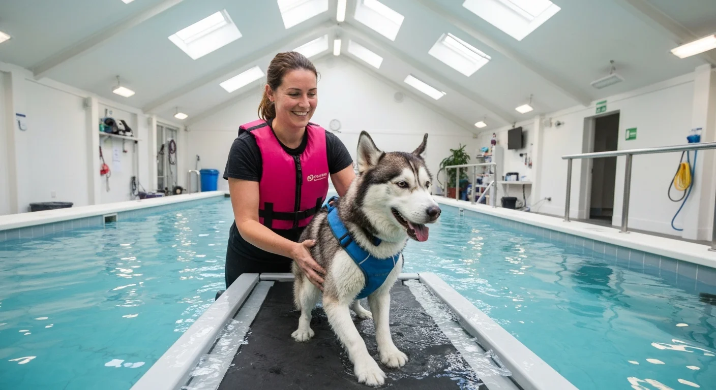
Hydrotherapy
Water-based exercise provides the most significant advantage for dogs with elbow dysplasia: substantial weight reduction through buoyancy while maintaining or increasing muscle activation.
| Modality | Weight Reduction | Primary Benefits | Considerations |
|---|---|---|---|
| Underwater treadmill (water at elbow) | 38-42% | Controlled gait pattern; adjustable speed and resistance; excellent for strengthening | Requires specialized equipment; session cost 40-80 GBP |
| Underwater treadmill (water at shoulder) | 58-62% | Maximum buoyancy support; suitable for early post-op or severely affected dogs | Reduced ground reaction forces may limit strengthening stimulus |
| Swimming (pool or open water) | ~100% (non-weight bearing) | Cardiovascular fitness; overall muscle conditioning; no joint loading | Cannot control gait pattern; some dogs resist water; requires supervision |
Underwater treadmill is generally preferred over swimming for elbow rehabilitation because it maintains a normal gait pattern and allows precise control of speed, duration, and resistance. Swimming, while excellent for cardiovascular conditioning, produces a paddling motion that does not replicate normal terrestrial gait and may actually increase elbow flexion-extension range beyond comfortable limits in some dogs.
Therapeutic Exercise
Structured exercises targeting specific muscle groups and movement patterns form the core of any rehabilitation program. The following exercises are commonly prescribed for dogs with elbow dysplasia:
Strengthening Exercises
- Controlled incline walking: 10-15 degree slopes target triceps and shoulder extensors
- Sit-to-stand transitions: Slow, controlled movements strengthen quadriceps and improve hindquarter support
- Three-leg standing: Lifting one limb forces increased loading on remaining limbs (use affected limb as standing limb)
- Resistance band walking: Elastic bands around the chest provide resistance during forward movement
Proprioception and Balance
- Wobble board standing: Unstable surface engages stabilizer muscles and improves joint position awareness
- Cavaletti poles: Low poles at measured intervals promote conscious limb placement and stride consistency
- Balance disc exercises: Progressive instability challenges coordination and core stability
- Figure-of-eight walking: Controlled turns at slow speed improve lateral stability and weight shifting
Manual Therapy
Hands-on techniques performed by trained rehabilitation practitioners address soft tissue restrictions, joint stiffness, and pain directly:
- Passive range of motion (PROM): Gentle movement of the elbow through its available range maintains joint mobility and provides feedback about stiffness progression.
- Joint mobilization: Skilled manual techniques applied to the elbow joint restore accessory gliding motions lost to capsular thickening and fibrosis.
- Soft tissue mobilization: Massage and myofascial release of compensatory muscle tension, particularly in the triceps, shoulder, and cervical musculature.
- Stretching: Controlled stretching of flexor and extensor muscle groups maintains functional range and reduces stiffness-related discomfort.
Physical Modalities
Various physical agents supplement exercise and manual therapy in managing ED-related pain and promoting tissue health:
| Modality | Mechanism | Evidence Level | Typical Application |
|---|---|---|---|
| Cryotherapy | Vasoconstriction; reduced nerve conduction; anti-inflammatory | Strong (well-established) | Post-exercise; acute flare management; 15-20 minutes |
| Thermotherapy | Vasodilation; increased tissue extensibility; muscle relaxation | Strong (well-established) | Pre-exercise; before manual therapy; 10-15 minutes |
| Laser therapy (LLLT/PBM) | Photobiomodulation; cellular energy production; anti-inflammatory | Moderate (growing evidence) | 2-3 sessions per week; direct application to joint |
| Therapeutic ultrasound | Deep tissue heating; improved blood flow; collagen remodeling | Limited in veterinary patients | Periarticular soft tissue; avoid over growth plates |
| TENS | Gate control pain modulation; endorphin release | Moderate | Pain management; can be applied at home with guidance |
| NMES | Direct muscle contraction via electrical stimulation | Moderate | Muscle strengthening when voluntary exercise insufficient |
Measuring Rehabilitation Outcomes
Objective outcome measurement distinguishes evidence-based rehabilitation from well-intentioned but unmeasured intervention. Several tools enable tracking of rehabilitation progress.
Objective Measures
- Force plate gait analysis: Measures peak vertical force and impulse for each limb during walking and trotting. The gold standard for detecting lameness and tracking recovery. A symmetry index comparing affected and sound limbs quantifies improvement objectively.
- Goniometry: Measurement of elbow range of motion using a goniometer. Normal canine elbow range is approximately 36 degrees flexion to 165 degrees extension. Serial measurements document stiffness progression or improvement.
- Thigh and forelimb circumference: Measured at standardized landmarks, limb circumference provides a proxy for muscle mass. Asymmetry between limbs indicates compensatory muscle changes.
- Body weight and condition score: Regular monitoring ensures weight management goals are maintained.
Subjective Measures
- Client-reported outcome measures: Validated questionnaires such as the Liverpool Osteoarthritis in Dogs (LOAD) and Canine Brief Pain Inventory (CBPI) track owner-perceived function and pain over time.
- Lameness scoring: Standardized visual lameness grading by a consistent observer provides reproducible assessment, though inter-observer variability limits precision.
Monitoring Frequency
For post-surgical rehabilitation, objective assessment every 2-4 weeks during the first 12 weeks allows timely protocol adjustments. For chronic management, quarterly assessments are sufficient for stable patients, with additional evaluation if clinical deterioration is suspected. Force plate data, when available, provides the most sensitive measure of subtle changes that visual assessment misses, similar to how CT scanning detects lesions invisible on radiographs during the diagnostic phase.
Home Exercise Programs
Clinic-based rehabilitation sessions typically occur once or twice weekly. The remaining days depend on owner-implemented home exercise programs (HEPs). The success of rehabilitation correlates strongly with HEP compliance, making program design and owner education critical components.
Designing Effective Home Programs
- Simplicity: Three to five exercises, clearly described, are more likely to be performed consistently than elaborate ten-exercise protocols.
- Demonstration: Every exercise should be demonstrated to the owner in person and ideally video-recorded for reference.
- Time-realistic: Total daily HEP time should not exceed 15-20 minutes. Programs requiring 45 minutes are abandoned within weeks.
- Progressive: Provide clear criteria for advancing exercises (e.g., "when your dog can balance on the wobble board for 30 seconds without stepping off, progress to the next level").
- Adaptable: Include "good day" and "bad day" versions of the program, acknowledging that osteoarthritis symptoms fluctuate.
Finding a Qualified Rehabilitation Practitioner
Canine rehabilitation is a specialized field requiring training beyond standard veterinary or physiotherapy qualifications. Several certification programs exist:
| Certification | Eligible Professionals | Recognized In |
|---|---|---|
| CCRT (Certified Canine Rehabilitation Therapist) | Veterinarians, physiotherapists, veterinary technicians | North America, internationally recognized |
| CCRP (Certified Canine Rehabilitation Practitioner) | Veterinarians, physiotherapists, veterinary technicians | North America, internationally recognized |
| ACVSMR (Diplomate, American College of Veterinary Sports Medicine and Rehabilitation) | Veterinarians (board certification) | North America |
| Chartered Physiotherapist (ACPAT Member) | Physiotherapists with animal therapy specialization | UK, Australia |
When selecting a rehabilitation provider, look for certification from one of these programs, experience with orthopedic cases (particularly elbow conditions), access to an underwater treadmill, and willingness to coordinate care with the referring veterinarian or surgeon. The best outcomes occur when the rehabilitation practitioner, surgeon, and primary care veterinarian communicate regularly about treatment goals and patient progress. For owners managing breeds predisposed to multiple joint health conditions, a rehabilitation practitioner experienced in comprehensive musculoskeletal management can address elbow, hip, and spinal issues within a unified treatment plan.
Related Database Resources
- Conservative Management - Non-surgical treatment framework
- Surgical Interventions - Procedures requiring post-operative rehabilitation
- ED in Working Dogs - Rehabilitation for return to duty
- Nutrition and Joint Development - Dietary support during recovery
Conclusion
Rehabilitation is not an optional luxury for dogs with elbow dysplasia; it is a core component of comprehensive management that measurably improves outcomes after surgery and maintains function in conservatively managed patients. The evidence supporting structured rehabilitation programs continues to accumulate, with post-surgical dogs showing faster recovery, better limb loading, and improved long-term function compared to rest-only protocols. For the veterinary community, the challenge is expanding access to qualified rehabilitation services and integrating rehabilitation planning into surgical discussions from the outset. For owners, understanding that surgery alone is not sufficient, and that their daily engagement with home exercise programs determines long-term outcomes, shifts expectations toward realistic, achievable goals in managing a progressive condition.