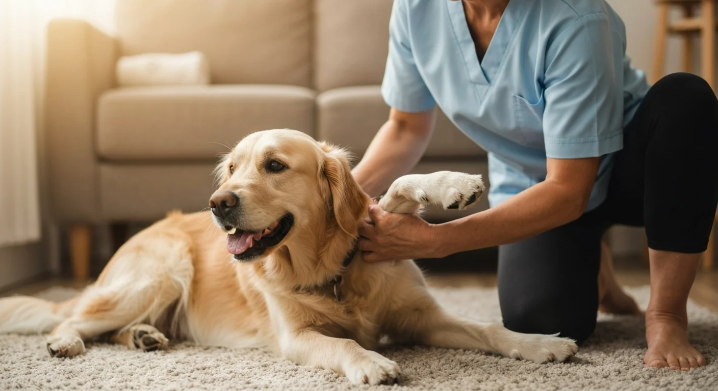
Secondary osteoarthritis is the inevitable long-term consequence of elbow dysplasia in virtually every affected dog. Regardless of whether the primary lesion is fragmented coronoid process, osteochondritis dissecans, or ununited anconeal process — and regardless of whether early surgical intervention has been performed — the initial cartilage damage and synovial inflammation set in motion a cascade of joint degeneration that progresses throughout the dog's life. Understanding this progression, monitoring it appropriately, and managing it comprehensively represents the long-game of elbow dysplasia treatment that extends years beyond the initial diagnosis and any surgical intervention.
Pathophysiology of OA Progression in Dysplastic Elbows
Osteoarthritis following elbow dysplasia is not simply the accumulation of wear on damaged cartilage. It is an active biological process driven by inflammatory mediators, enzymatic cartilage degradation, and subchondral bone remodeling. Once the initial dysplastic lesion disrupts joint congruence and cartilage integrity, a self-perpetuating cycle of inflammation and degeneration establishes itself that modern medicine can slow but not halt.
The key mediators of OA progression in dysplastic elbows include interleukin-1 beta (IL-1β) and tumor necrosis factor-alpha (TNF-α), which stimulate chondrocytes to produce matrix metalloproteinases (MMPs). These enzymes degrade collagen and proteoglycans — the structural proteins of articular cartilage — faster than chondrocytes can synthesize replacements. As the cartilage matrix degrades, synovial fluid loses its lubricating and shock-absorbing properties, accelerating mechanical wear. Synovial membrane hypertrophy and villous transformation contribute to chronic joint effusion, while subchondral bone eburnation (sclerosis and hardening) occurs in response to increased loading of denuded bone surfaces.
OA Progression Rates
Longitudinal studies tracking OA progression in dogs with elbow dysplasia consistently find measurable radiographic change even in dogs managed surgically and with optimal aftercare. Fitzpatrick et al. (2009) followed 53 dogs with FCP treated arthroscopically over 36 months, finding progressive osteophyte formation in 87% of cases despite successful fragment removal. The rate of progression correlated most strongly with the extent of cartilage damage present at surgery — emphasizing that early intervention, before significant cartilage destruction accumulates, meaningfully influences the long-term OA trajectory even if it cannot prevent progression entirely.
Radiographic Progression: What to Monitor
Serial radiographic evaluation allows objective monitoring of OA progression over time, guiding treatment intensification decisions before clinical deterioration becomes obvious. The following radiographic features are assessed and graded at follow-up evaluations:
| Radiographic Feature | Location | Clinical Significance | Assessment Interval |
|---|---|---|---|
| Osteophyte formation | Caudal humeral condyle; anconeal process; medial coronoid | Primary OA marker; size correlates loosely with pain burden | Annual |
| Subchondral sclerosis | Ulnar notch; humeral trochlea | Indicates bone remodeling under increased loading | Annual to biennial |
| Joint space narrowing | Overall elbow joint | Reflects cartilage loss; advanced sign | Biennial |
| Periarticular mineralization | Soft tissue adjacent to joint | Advanced OA change; may impinge on soft tissue | As clinically indicated |
Establishing a radiographic baseline at the time of initial diagnosis or surgical intervention provides essential reference data for follow-up comparisons. Radiographs taken years later without baseline images make it impossible to quantify progression rate, which is often more clinically relevant than absolute OA grade at a single time point.
Clinical Staging of Elbow OA
Functional OA staging guides treatment intensity. The following clinical staging framework, adapted from the WSAVA pain management guidelines, provides a practical approach:
Stage 1 — Subclinical
Radiographic OA without consistent clinical signs. Dog maintains normal activity. Management: weight optimization, annual monitoring, owner education about progression signs, omega-3 supplementation, consider low-intensity hydrotherapy.
Stage 2 — Mild
Intermittent lameness after exercise; stiffness after rest. Performance reduction noted by owners. Management: NSAID therapy for flares, regular physical rehabilitation, activity modification, weight management intensification.
Stage 3 — Moderate
Persistent lameness; visible gait abnormality at rest and exercise. Reduced quality of life. Management: continuous NSAID therapy, multimodal analgesia with adjunct drugs, regular rehabilitation, consideration of advanced modalities (laser, TENS).
Stage 4 — Severe
Severe persistent lameness; significantly restricted mobility; sleep disturbance from pain. Management: maximal multimodal analgesia, intensive rehabilitation, consideration of total elbow replacement, quality of life assessment.
Medical Management of OA in Dysplastic Elbows
The principles of managing secondary OA in elbow dysplasia align with those for OA at any joint, but the elbow's complex tri-osseous articulation and the specific pattern of medial compartment disease create some specific management considerations.
NSAID Therapy
Non-steroidal anti-inflammatory drugs remain the pharmacological cornerstone of OA management. For dogs with persistent clinical signs at Stage 2 or above, daily NSAID administration provides superior outcomes compared to intermittent dosing at flares. Clinical response should be formally assessed using validated pain scales (CBPI, LOAD, or similar) at 4-week intervals after initiation, and NSAID choice should be revisited if adequate response is not achieved within 4-6 weeks. As detailed in the pain management guide, only veterinary-approved NSAIDs should be used, with appropriate safety monitoring.
Disease-Modifying Osteoarthritic Agents
Several categories of agents are used with chondroprotective intent, though the strength of evidence varies considerably:
- Omega-3 fatty acids (EPA/DHA): The strongest evidence base among supplements. A meta-analysis by Roush et al. (2010) demonstrated significant improvement in force plate measurements and clinical scores in dogs with OA receiving therapeutic doses of fish oil (EPA+DHA ≥75 mg/kg/day). Anti-inflammatory through 5-lipoxygenase pathway modulation.
- Pentosan polysulfate: Inhibits protease enzymes that degrade cartilage matrix; reduces synovial inflammation. Administered as injections rather than oral supplementation for bioavailability reasons. Typically 3-4 mg/kg subcutaneously weekly for four weeks, then monthly maintenance.
- Polysulfated glycosaminoglycans (Adequan): Provides cartilage matrix precursors and inhibits degradative enzymes. Standard protocol: 4.4 mg/kg IM biweekly for 8 injections, then monthly maintenance. Evidence quality for cartilage protection is moderate in dogs.
- Joint supplements (glucosamine/chondroitin): Despite widespread use, clinical trial evidence in dogs is inconsistent. Some controlled trials show modest benefit; others show no significant effect versus placebo. These supplements are unlikely to harm, but should not substitute for evidence-based therapies when clinical signs are present.
Newer Biological Therapies
Monoclonal antibody therapies targeting nerve growth factor (NGF), similar to frunevetmab approved in cats and under investigation in dogs, represent a new class of OA analgesics that may become available for canine elbow OA within the next few years. These biologics target pain pathways without the GI and renal risks associated with NSAIDs, potentially enabling better long-term pain control in dogs with NSAID intolerance or concurrent renal disease.
Platelet-rich plasma (PRP) and mesenchymal stem cell (MSC) therapies are available at specialist centers, with an evolving evidence base in canine OA. Intra-articular PRP injections have shown promising results in several clinical trials, reducing inflammatory markers in synovial fluid and improving short-term force plate measurements. However, optimal protocols, long-term efficacy, and cost-effectiveness compared to existing therapies remain under investigation.
Physical Rehabilitation for Chronic OA
Long-term physical rehabilitation is an underutilized component of OA management in dysplastic elbows. The goals evolve from the acute post-surgical phase to long-term maintenance: preserving range of motion against progressive capsular fibrosis, maintaining periarticular muscle mass to share joint loading, and providing pain relief through physical modalities. The rehabilitation and physical therapy article covers exercise protocols in detail; for OA management specifically, the key interventions are:
- Hydrotherapy: Warm water reduces pain perception and allows muscle maintenance without full weight-bearing. Monthly or bimonthly hydrotherapy sessions for Stage 2-3 OA dogs maintain function between exacerbation periods.
- Therapeutic laser (Class IV): Reduces inflammatory cytokine expression and promotes cellular healing in chronically inflamed synovium. Evidence for laser therapy in canine elbow OA has strengthened considerably since 2018.
- Massage and passive range of motion: Regular home-based passive elbow flexion-extension within comfortable range prevents capsular fibrosis progression. Owners can be taught appropriate techniques with professional guidance.
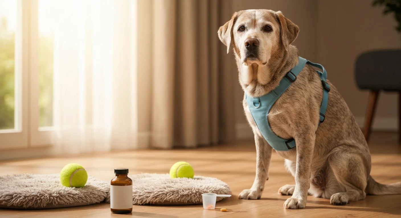
Weight Management: The Most Impactful Intervention
No single management intervention has a larger evidence base for improving OA outcomes than weight management. The mechanics are straightforward: at a trot, the elbow joint bears approximately 1.5-2 times body weight per loading cycle. In a 35 kg Labrador Retriever, a 5 kg reduction in body weight — approximately 14% — reduces elbow joint loading by 7.5-10 kg per stride. Across thousands of daily steps, this reduction substantially decreases cumulative joint stress and inflammatory load. A landmark study by Impellizeri et al. (2000) demonstrated that weight loss alone produced OA improvement equivalent to NSAID therapy in overweight dogs — without pharmacological risk.
Weight Management Requires Active Veterinary Monitoring
Weight loss in dogs with chronic OA must be managed carefully to prevent concurrent muscle wasting. A combination of caloric restriction with maintained protein intake, combined with low-impact exercise to preserve muscle mass during weight loss, produces better outcomes than caloric restriction alone. Target lean body condition (4/9 BCS) rather than minimum weight, and reassess body condition monthly during weight loss programs.
Related Database Resources
- Pain Management in Elbow Dysplasia - Analgesic protocols for OA pain
- Rehabilitation and Physical Therapy - Exercise programs for chronic OA
- Conservative Management - Non-surgical treatment overview
- Surgical Interventions - When surgery influences OA trajectory
Conclusion
Secondary osteoarthritis is the unavoidable long-term reality of elbow dysplasia. It begins with the initial joint injury and progresses throughout the dog's life, but its rate of progression and its impact on quality of life are substantially modifiable through evidence-based management. The framework of OA staging, multimodal analgesia, regular physical rehabilitation, and consistent weight management — applied proactively rather than reactively — is the foundation of good long-term care for every elbow dysplasia patient. Dogs diagnosed early and managed comprehensively from diagnosis onward achieve far better lifelong quality of life than those who receive treatment only when OA has become clinically obvious.