Elbow incongruity — the imperfect geometric fit between the three bones of the canine elbow joint — occupies a contested but increasingly central position in the understanding of elbow dysplasia pathogenesis. Long considered a secondary consequence of developmental abnormalities, accumulating evidence suggests that incongruity may in many cases be the primary mechanical driver that initiates fragmented coronoid process formation, contributes to ununited anconeal process failure, and accelerates secondary osteoarthritis development. Whether incongruity is classified as a fourth component of elbow dysplasia or as the unifying mechanism underlying all three classical components, clinicians who understand its biomechanical basis and diagnostic features will manage elbow dysplasia patients more effectively than those who do not.
The Anatomy of Congruence
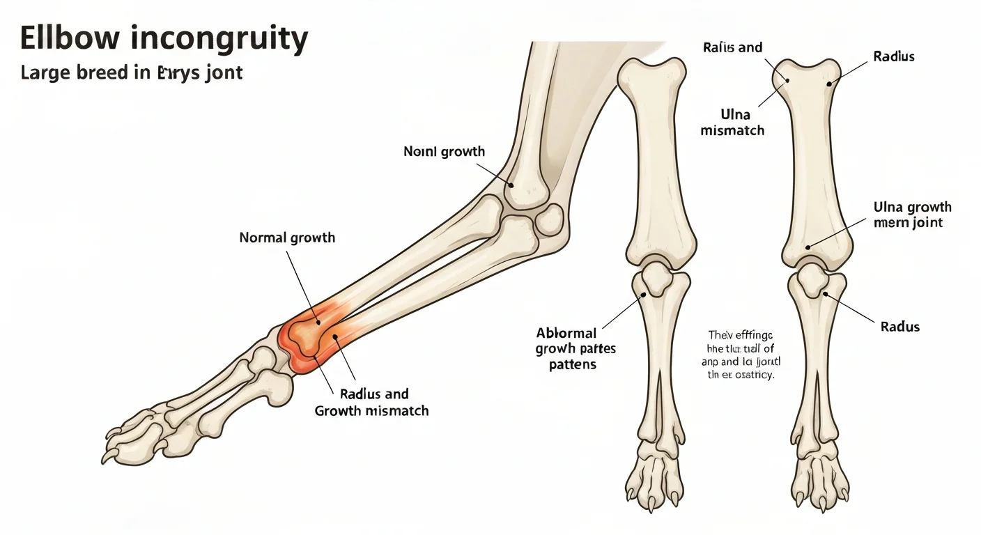
The canine elbow joint is one of the most geometrically constrained joints in veterinary medicine. Three bones — the humerus, radius, and ulna — must articulate with submillimeter precision to distribute joint loading forces evenly across the entire articular surface. The humeral trochlea sits within the trochlear notch of the ulna, while the humeral capitulum articulates with the radial head. These contact points are not independent; they function as a unified system where the relative position of radius and ulna determines load distribution across both articular surfaces simultaneously.
During development, the radius and ulna must grow at precisely synchronized rates. If the radius is proportionally shorter than the ulna (short radius incongruity), the humeral capitulum cannot engage the radial head at the same depth as the trochlea engages the ulnar notch. This creates a "step" in the joint surface at the radioulnar junction, concentrating contact stress on the caudal humeral condyle. Conversely, if the ulna is proportionally shorter (short ulna incongruity, less common), the anconeal process bears disproportionate load as the trochlea overrides its caudal surface during joint flexion.
How Small Is Too Small?
Experimental studies by Preston et al. (2001) used cadaveric elbow specimens with controlled radio-ulnar length discrepancy to quantify the mechanical consequences of incongruity. A 1.5 mm short radius incongruity increased contact pressure on the medial coronoid process by 47% compared to the congruent elbow. A 3 mm discrepancy tripled medial coronoid pressure. Given that the medial coronoid is the most common site of FCP fragmentation, these findings provide a direct mechanical explanation for why even modest incongruity dramatically increases FCP risk in genetically predisposed individuals.
Classification of Elbow Incongruity
Three distinct incongruity types have been described, each with different mechanical consequences and clinical associations:
Type 1: Short Radius Incongruity
The most common type. The radial head sits proximal to its ideal position within the trochlear notch. Creates a "step" at the radiohumeral junction that overloads the medial coronoid process. Strongly associated with FCP. Most common in Labrador and Golden Retrievers.
Type 2: Short Ulna Incongruity
Less common. The ulna is short relative to the radius; the trochlea bears against the anconeal process abnormally during flexion. Associated with UAP in some cases. Can occur secondary to premature closure of the distal ulnar physis.
Type 3: Elliptical Trochlear Notch
The geometry of the ulnar trochlear notch is ovoid rather than circular, creating incomplete articulation with the round humeral trochlea. This form of incongruity does not involve length discrepancy but creates inherently non-uniform contact pressure distribution throughout the range of motion.
Relationship to FCP, UAP, and OCD
The clinical significance of incongruity lies primarily in its relationship to the classical elbow dysplasia components. The precise causal relationships remain debated, but the weight of evidence supports the following associations:
Incongruity and FCP
Short radius incongruity and type 3 trochlear notch incongruity are detected in high proportions of FCP cases. Samoy et al. (2006) found measurable incongruity in 62% of FCP cases examined by CT, compared to 12% of normal control elbows. The medial coronoid process, when subjected to abnormal contact pressure from incongruity, undergoes fatigue loading that eventually results in fissuring and fragmentation. This mechanically-derived explanation for FCP has largely supplanted earlier theories involving pure osteochondrosis of the coronoid tip.
Incongruity and UAP
The anconeal process normally fuses with the olecranon by 4-5 months of age in large breeds. This fusion is dependent on compressive stress across the growth plate during normal weight-bearing, which promotes ossification. When short ulna incongruity creates abnormal shear rather than compressive loading across the anconeal physis during the critical fusion window, fusion may fail — resulting in ununited anconeal process. This incongruity-dependent model explains why UAP has a different breed and age distribution than FCP and why ulnar osteotomy can allow late fusion of some UAP cases.
Incongruity and OCD
The relationship between incongruity and osteochondritis dissecans of the medial humeral condyle is less direct than for FCP. OCD is primarily an osteochondrosis lesion — a failure of endochondral ossification — rather than a mechanically-induced fracture. However, incongruity-associated changes in contact stress distribution may influence which areas of the medial humeral condyle develop OCD lesions by creating focal zones of abnormally high compressive loading in a cartilage that may be already predisposed to osteochondrosis.
Diagnosis of Elbow Incongruity
Accurately diagnosing and quantifying elbow incongruity requires appropriate imaging. Standard radiographs have limited sensitivity for detecting subtle incongruity — the two-dimensional projection of a three-dimensional joint geometry creates significant information loss.
Radiographic Assessment
The mediolateral flexed view allows assessment of the step between the radial head and the trochlear notch at the level of the radioulnar junction. A clearly visible step of 1 mm or more is a radiographically detectable sign of significant short radius incongruity. However, positioning variation, magnification differences, and projection angle significantly affect the apparent step height, making precise radiographic quantification unreliable for steps smaller than 2 mm.
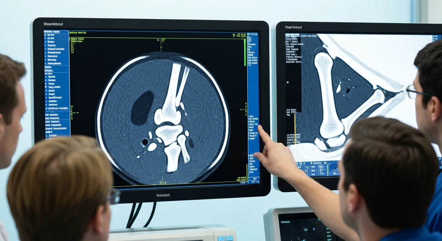
CT Assessment
Computed tomography is the gold standard for incongruity assessment, providing cross-sectional images that directly visualize the relationship between the radial head, ulnar trochlear notch, and humeral condyles in multiple planes. CT scanning allows precise measurement of radio-ulnar step height in standardized planes, identification of the incongruity type, and simultaneous assessment for concurrent FCP or UAP. For dogs in which surgical correction of incongruity is contemplated, CT measurements are essential for pre-operative planning.
Arthroscopic Assessment
Diagnostic arthroscopy provides direct visualization of the radiohumeral and trochlear joints, allowing real-time assessment of articular contact under approximately physiological loading. The "palpation test" during arthroscopy — probing the radioulnar junction with a blunt instrument to assess step height — correlates reasonably well with CT measurements and adds the advantage of direct cartilage assessment in the same procedure. Most dogs undergoing arthroscopic FCP treatment have concurrent incongruity assessment performed at the same time.
Surgical Treatment of Incongruity: Proximal Ulnar Osteotomy
For dogs with confirmed significant incongruity (typically radioulnar step of 2 mm or more) contributing to FCP, dynamic proximal ulnar osteotomy (PUO) or distraction osteogenesis (lengthening) of the radius have been developed to restore joint congruence and reduce abnormal coronoid loading.
Proximal Ulnar Osteotomy Technique
PUO involves a controlled osteotomy of the proximal ulna, proximal to the coronoid process. By dividing the ulna, the mechanical constraint between radius and ulna is released. The relatively elastic soft tissue structures then allow the radius and ulna to adopt a more congruent relationship under the natural joint contact forces from the humeral condyle. Ideally, the radial head "seats" more deeply against the capitulum, reducing the step at the radiohumeral junction and the abnormal coronoid loading.
The procedure is typically performed arthroscopically-assisted, with FCP fragment debridement performed first, followed by PUO through a separate small incision. The osteotomy gap is left open to heal by secondary intention — the biological process of bone bridging across the gap takes 6-12 weeks and is monitored radiographically.
Evidence for PUO
The evidence base for PUO has expanded significantly since 2010. Fitzpatrick et al. (2009) and Cook et al. (2012) both found that dogs with radioulnar incongruity treated with concurrent PUO and FCP debridement showed superior long-term force plate outcomes compared to FCP debridement alone in incongruent elbows. However, outcomes in elbows with minimal incongruity did not benefit from PUO addition. These findings support selective use of PUO in confirmed significant incongruity rather than routine addition to all FCP procedures.
Conservative Management of Incongruity
Not all dogs with elbow incongruity are candidates for or owners of corrective surgery. For older dogs with established OA, very mild incongruity, or owner constraints limiting surgical options, conservative management targeting the consequences of incongruity — FCP-associated pain, coronoid OA, and progressive joint degeneration — follows the same protocols as general elbow dysplasia conservative management. Weight optimization to reduce joint loading is particularly important in incongruent elbows where each kilogram of body weight is amplified by the mechanical disadvantage of non-congruent joint geometry.
Screening Implications
Elbow incongruity is not formally graded by the IEWG, OFA, or BVA/KC certification schemes — these schemes grade consequences (osteophytosis, primary lesions) rather than causes. However, some progressive registries are beginning to include incongruity notation on screening reports, reflecting growing recognition of its clinical significance. For breeders, awareness that dogs with lower-grade OA changes but visible incongruity on radiographs may have higher breeding risk than the grade alone suggests represents an evolving refinement of how to interpret screening data.
Related Database Resources
- FCP: Fragmented Coronoid Process - Incongruity as a primary FCP driver
- UAP: Ununited Anconeal Process - Short ulna incongruity relationship
- CT Scanning for Elbow Dysplasia - Gold standard incongruity assessment
- Surgical Interventions - PUO in context of other ED procedures
Conclusion
Elbow incongruity represents one of the most mechanistically compelling explanations for how elbow dysplasia components arise and why they progress despite treatment. The accumulating evidence that even modest radio-ulnar length discrepancy substantially increases medial coronoid loading supports a biomechanical rather than purely osteochondrotic model of FCP pathogenesis in most cases. For clinicians, recognizing incongruity through appropriate imaging — CT for quantification, arthroscopy for direct visualization — allows treatment planning that addresses the mechanical cause rather than only its structural consequences. For breeders, understanding that some degree of incongruity may be heritable suggests that detailed pedigree analysis of FCP prevalence in lines with confirmed incongruity warrants similar scrutiny to FCP prevalence alone.