Elbow dysplasia is fundamentally a disease of development. The structural abnormalities that define the condition — fragmented coronoid process, osteochondritis dissecans of the medial humeral condyle, and ununited anconeal process — develop during the explosive growth phase between four and eight months of age. This developmental window represents both the period of maximum vulnerability and, critically, the period of greatest opportunity for intervention. For breeders and puppy owners of susceptible large breeds, understanding the early clinical signs of elbow dysplasia, knowing when to seek veterinary evaluation, and appreciating how early diagnosis changes outcomes can meaningfully improve welfare in predisposed lines.
The Developmental Window: Why Timing Matters
The canine elbow joint is a complex tri-osseous articulation between the humerus, radius, and ulna. During rapid growth, these three bones must grow synchronously to maintain joint congruence. Asynchronous growth, even by 1-2 millimeters of radio-ulnar length discrepancy, creates abnormal pressure distribution across the developing joint surfaces. The medial coronoid process of the ulna, which bears approximately 60% of the elbow's medial compartment load, is particularly vulnerable to this incongruity-driven overloading.
The ossification centers of the canine elbow close sequentially between three and five months of age, with the anconeal process fusing with the ulna between four and five months in large breeds. When fusion fails — the defining event of ununited anconeal process — it is this developmental window that has been disrupted. Similarly, the fragmentation or fissuring of the medial coronoid process occurs during peak loading on a joint that has not yet fully matured.
Why Early Signs May Be Subtle
Puppies are extraordinarily compensatory. Unlike adult dogs with established movement patterns, puppies naturally adjust their gait as they learn to move, making subtle asymmetry difficult to detect without trained observation. Additionally, bilateral elbow dysplasia — which occurs in 50-60% of affected dogs — creates a symmetrically abnormal gait that owners frequently attribute to "puppy clumsiness" rather than joint disease. Studies by Kirberger (2007) found that owners reported onset of noticed signs at 6.2 months on average, while radiographic evidence suggested lesions had been developing since 4-5 months.
Clinical Signs by Age Group
Four to Six Months: Earliest Indicators
The earliest signs of elbow dysplasia at this age are often behavioral rather than overtly orthopedic. Affected puppies may show reluctance to engage in play that requires repetitive elbow loading — fetching, wrestling with littermates, stair climbing. Owners sometimes report that the puppy seems less engaged, tires more quickly on walks, or occasionally holds one foreleg slightly off the ground after exertion.
- Stiffness after rest: The puppy rises slowly and takes several steps to "warm up" before moving normally. This is a classic early sign that is frequently dismissed as normal puppy behavior.
- Intermittent forelimb lifting: Lifting one front leg when standing still, particularly after exercise, indicates elbow discomfort. This sign is more obvious when unilateral disease is present.
- Reduced play tolerance: Preferring to stop and rest during play sessions that littermates continue without difficulty.
- Protective posturing: Sitting with affected leg extended rather than flexed, or lying with elbow in an unusual position.
Six to Ten Months: More Obvious Presentation
As the puppy grows heavier and joint damage progresses, clinical signs typically become more apparent. This is the age range at which most puppies are first presented to veterinarians for elbow dysplasia evaluation. By this stage, significant secondary joint changes may have already begun.
- Observable lameness: A visible head bob or shortened stride on the affected leg, worsening during or after exercise.
- Joint swelling: Visible or palpable effusion around the elbow joint, particularly on the medial aspect.
- Elbow held outward (abduction): Dogs with medial compartment pain often carry the affected elbow slightly abducted to reduce medial joint contact pressure. This creates a characteristic "paddling" front limb movement at trot.
- Reduced range of motion: Reluctance or inability to fully flex or extend the elbow compared to the opposite limb.
- Pain on manipulation: Vocalization or withdrawal when the elbow is flexed under gentle pressure.
FCP Presentation
Fragmented coronoid process typically presents at 5-9 months. Medial elbow pain predominates; dogs often show lameness that improves after 10-15 minutes of movement. Bilateral presentation is common (60-70% of cases), potentially masking obvious asymmetric lameness.
UAP Presentation
Ununited anconeal process classically presents at 4-7 months, often with more acute severe lameness than FCP. The anconeal process fails to fuse; its mechanical instability causes acute joint pain. German Shepherds and Saint Bernards show highest UAP prevalence.
OCD Presentation
Osteochondritis dissecans of the medial humeral condyle typically presents at 6-10 months. Lameness may be severe if the cartilage flap detaches as a "joint mouse." Labrador and Golden Retrievers show relatively higher OCD rates compared to FCP.
When to Seek Veterinary Evaluation
Any large breed puppy showing forelimb lameness, stiffness after rest, reluctance to exercise, or joint swelling should be evaluated by a veterinarian promptly. Early evaluation is preferable to waiting for signs to become more obvious — by the time lameness is severe and persistent, secondary joint changes are typically already underway. Specific triggers for immediate evaluation include:
- Forelimb lameness lasting more than 24-48 hours in a large breed puppy under 12 months
- Acute onset of severe lameness in a large breed puppy (weight-bearing lameness becoming non-weight-bearing)
- Visible joint swelling around the elbow
- Consistent stiffness after rest across multiple episodes
- Puppy from a breed with elevated ED prevalence (German Shepherd, Labrador, Rottweiler, Bernese Mountain Dog) showing any forelimb abnormality
Breed Risk Context
The threshold for seeking evaluation should be lower in high-risk breeds. A six-month Labrador Retriever from a line without documented elbow screening that shows intermittent forelimb stiffness has a meaningfully higher probability of elbow dysplasia than a Miniature Poodle with similar signs. Breed-specific prevalence data provides the risk context that informs triage decisions.
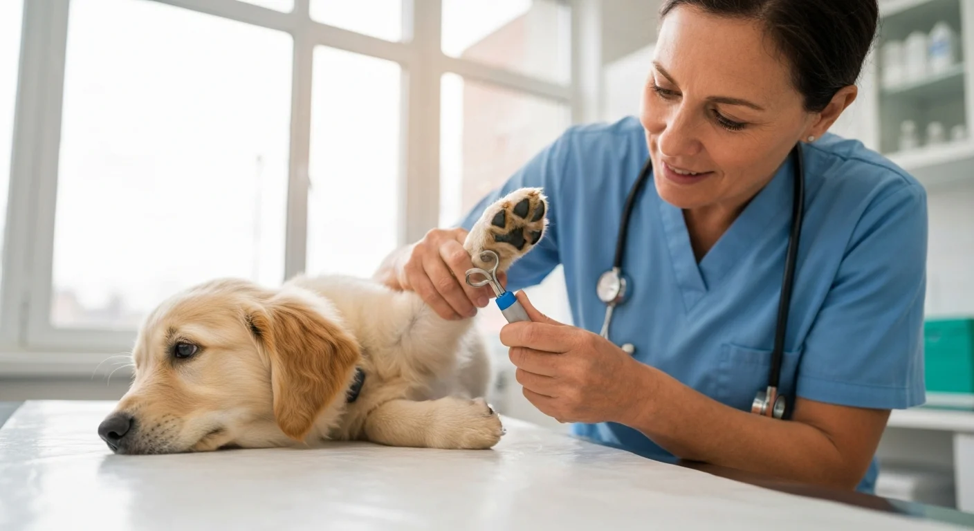
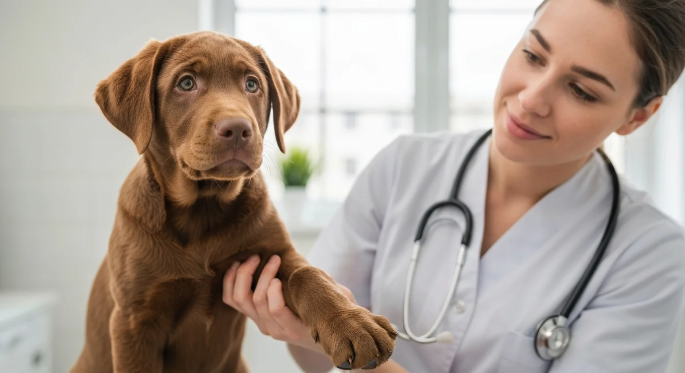
Veterinary Evaluation in Young Dogs
Physical examination of a puppy with suspected elbow dysplasia should include systematic joint palpation, range of motion assessment, and gait evaluation at walk and trot. The "coronoid compression test" — applying medial pressure while cycling through elbow flexion and extension — elicits pain in most dogs with active FCP. However, physical examination findings alone cannot differentiate ED components or confirm the diagnosis definitively.
Radiographic Evaluation in Puppies
Standard radiographic views in young dogs with suspected ED include mediolateral (flexed) and craniocaudal projections. In very young puppies (under five months), normal developmental ossification patterns can mimic or obscure pathological changes, and radiographic interpretation requires an experienced veterinary radiologist or orthopedic specialist.
The anconeal process should be visible as a separate ossification center until 4-5 months of age; failure to fuse by five months in large breeds, or by four months in German Shepherds, supports a UAP diagnosis. Fragmented coronoid process is notoriously difficult to visualize on plain radiography — sensitivity is only 60-70% — which is why CT scanning is often recommended when radiographs are equivocal in young dogs with clear clinical signs.
Early Intervention: Does It Improve Outcomes?
The evidence for early surgical intervention improving long-term outcomes in elbow dysplasia is strongest for ununited anconeal process and moderately strong for fragmented coronoid process. The key principle is limiting secondary osteoarthritis development: every week that an unstable or fragmented lesion remains in the joint contributes to progressive cartilage damage and synovial inflammation that cannot be reversed.
A retrospective study by Huibregtse et al. (2001) compared UAP outcomes in dogs operated before versus after six months of age. Dogs operated before six months showed significantly lower osteoarthritis scores at two-year follow-up and higher return to normal function rates. Similar findings have been replicated in multiple studies since, establishing early surgical intervention for UAP as the standard of care when the diagnosis is confirmed before skeletal maturity.
For FCP, the outcome advantage of early surgery is less absolute, partly because FCP-associated cartilage damage begins before fragments are fully formed and partly because the extent of concurrent cartilage damage is not fully predictable preoperatively. Nevertheless, arthroscopic removal of FCP fragments performed before advanced secondary OA develops consistently produces better long-term outcomes than late intervention.
Management While Awaiting Diagnosis
While awaiting veterinary evaluation or diagnostic imaging, the following measures minimize joint damage and maintain puppy welfare:
- Restrict high-impact exercise: No running off-lead, no jumping, no stairs. Leash walks on soft surfaces only.
- Maintain body weight at lean: Reducing mechanical joint loading by keeping the puppy lean may reduce symptom progression. Avoid overfeeding to "support growth."
- Avoid calcium supplementation: If not already established, there is no benefit to supplementation and potential harm from disrupting endochondral ossification.
- Soft sleeping surface: Orthopaedic foam or padded bedding reduces pressure on affected joints during rest periods.
- Pain management only under veterinary guidance: Never administer human NSAIDs to a puppy. Veterinary NSAIDs at appropriate doses may be prescribed while awaiting specialist evaluation.
Related Database Resources
- Elbow Dysplasia Overview - Comprehensive background on the condition
- FCP: Fragmented Coronoid Process - Most common ED component in detail
- UAP: Ununited Anconeal Process - Surgical emergency in young dogs
- Screening Protocol - Certification screening at maturity
Breeder Responsibilities
Breeders play a critical role in early detection. Litters from lines with known ED prevalence should be observed systematically for forelimb gait abnormalities during the 4-12 month growth period. Puppy contracts that include provisions for veterinary evaluation of any lameness, combined with breeder transparency about parental screening results, create a framework for early intervention that serves both puppy welfare and the long-term health of the line. Breeders who implement evidence-based breeding decisions using elbow scores from both parents and extended family members reduce, though cannot eliminate, ED risk in offspring from high-prevalence lines.
Conclusion
Elbow dysplasia in puppies is detectable before significant secondary damage accumulates, but only if owners and breeders know what to look for and act promptly when early signs appear. The four-to-twelve-month developmental window is simultaneously the period of greatest disease formation and greatest treatment opportunity. For large breed puppies from predisposed lines, a low threshold for veterinary evaluation of any forelimb abnormality, combined with appropriate environmental management during growth, represents the most effective approach to limiting the long-term impact of this common heritable condition.