Elbow dysplasia is most treatable when it is caught early. Yet the condition remains chronically under-diagnosed in its initial stages because the clinical signs in growing dogs are subtle, intermittent, and easily attributed to normal puppy clumsiness. By the time lameness becomes obvious and persistent, significant cartilage damage has often already occurred. Understanding when and how elbow dysplasia first manifests allows breeders, owners, and veterinarians to intervene during the narrow window where treatment outcomes are substantially better. This article synthesizes the current evidence on age of onset, early clinical indicators, and the examination techniques that improve detection rates before irreversible joint changes develop.

The Critical Window: When Elbow Dysplasia Develops
Elbow dysplasia is not a single disease but a group of developmental conditions that share a common origin in abnormal growth of the elbow joint during skeletal maturation. The three primary components, fragmented coronoid process (FCP), osteochondritis dissecans (OCD), and ununited anconeal process (UAP), each have somewhat different developmental timelines, but all originate during the rapid growth phase between approximately 4 and 10 months of age.
The radius and ulna grow at slightly different rates during this period. In a normally developing elbow, the growth plates of both bones coordinate precisely so that the joint surfaces remain congruent. When this synchrony is disrupted, whether by genetic predisposition, nutritional factors, or biomechanical stress, the resulting joint incongruity creates abnormal pressure distribution across the articular cartilage and subchondral bone. It is this incongruity that initiates the pathological cascade leading to fragmentation, cartilage flap formation, or failure of the anconeal process to fuse.
Age of First Clinical Signs by ED Component
FCP: Clinical signs typically emerge between 5 and 9 months, though some dogs do not show obvious lameness until 12-18 months when secondary osteoarthritis develops. FCP is the most common component and the most frequently missed early because lameness is often bilateral and symmetrical.
OCD: Signs generally appear between 5 and 8 months, coinciding with the period of maximum endochondral ossification in the medial humeral condyle. Lameness may be acute if a cartilage flap detaches.
UAP: The anconeal process normally fuses to the ulna by 16-20 weeks (breed-dependent). Failure of fusion becomes radiographically apparent after 20 weeks, though clinical signs may be delayed until secondary instability and OA develop, often around 6-12 months.
Why Early Signs Are So Easily Missed
Several characteristics of early elbow dysplasia conspire to delay diagnosis. Understanding these patterns helps explain why so many dogs are not identified until the disease is well established.
Bilateral Disease Masks Lameness
Elbow dysplasia is bilateral in approximately 50-70% of affected dogs, depending on the breed and the specific component involved. When both elbows are similarly affected, the dog does not develop the characteristic asymmetric gait that owners and veterinarians associate with lameness. Instead, the dog may show a shortened stride on both forelimbs, reduced willingness to play, or subtle changes in posture, none of which scream "orthopedic problem" to most observers. The absence of a clear head bob or weight-shifting limp leads owners to describe the dog as "a bit lazy" or "not as active as his littermates" rather than suspecting a developmental joint condition.
Intermittent Presentation
Early ED lameness is classically intermittent. Dogs may be stiff after rest (particularly after prolonged sleep), improve with gentle movement, then worsen again after vigorous exercise. This waxing and waning pattern leads owners to rationalize: the dog "slept funny," "played too hard," or "tweaked something" and is now better. The resolution of lameness between episodes provides false reassurance that nothing serious is wrong.
Puppy Resilience and Drive
Young dogs, particularly those of high-drive working breeds that are most susceptible to ED, have remarkable capacity to compensate for joint pain. Their enthusiasm for activity and social interaction can override pain signals that would clearly limit an older dog. A 7-month-old German Shepherd with developing FCP will still chase a ball, still wrestle with housemates, still bound to the door when the leash appears. The pain is present, but so is the motivation to ignore it.
Gradual Onset
Because ED develops progressively rather than from a single traumatic event, there is no sudden before-and-after transition that would alert an observer. The dog was never "normal and then lame." Instead, the condition insidiously progresses from subclinical to mildly symptomatic to obviously lame over weeks or months, making it difficult to identify a definitive onset.
Early Clinical Signs: What to Watch For
Identifying ED before significant cartilage damage occurs requires attention to subtle indicators that precede obvious lameness. The following signs, particularly in combination, should raise clinical suspicion in breeds with known ED predisposition.
Gait Abnormalities
- Shortened forelimb stride: The most consistent early gait change. The dog takes shorter steps with the front legs compared to the rear, reducing elbow flexion and extension. This is easiest to observe when watching from the side at a walk or slow trot.
- Stiffness after rest: Often most pronounced first thing in the morning or after napping. The dog may be reluctant to rise, move gingerly for the first 5-10 minutes, then gradually warm up. Owners typically notice this resolves quickly and dismiss it.
- Reluctance to descend stairs: Descending stairs loads the forelimbs disproportionately. Dogs with early ED may hesitate at the top of stairs, descend slowly, or avoid stairs they previously navigated without concern.
- Sitting abnormally: Some dogs with bilateral forelimb discomfort shift to sitting with their front legs splayed outward (externally rotated), spreading weight across the lateral compartment and reducing medial elbow loading.
- Paddling or winging gait: Some affected dogs develop a characteristic outward swing of the forepaw during the swing phase. This circumduction reduces elbow flexion and the associated pain during the stride cycle.
Behavioral Changes
- Decreased play tolerance: The dog may initiate play but tire quickly, lie down during sessions, or become irritable when housemates continue to roughhouse.
- Reluctance to jump: Previously enthusiastic jumpers (onto furniture, into vehicles) may hesitate, require encouragement, or find alternative routes that avoid jumping.
- Activity-dependent stiffness: Lameness or stiffness that consistently appears 1-2 hours after vigorous exercise, even if absent during the activity itself.
- Changed lying position: Dogs may avoid fully extending their forelimbs when lying down, preferring to tuck them under the body to reduce elbow extension.
- Weight shifting: Subtle alternation of weight between forelimbs while standing, visible as a gentle rocking motion when observed from behind.
Breed Context Matters
These signs carry far greater significance in breeds with established ED predisposition. A 6-month-old Labrador Retriever or Rottweiler showing morning stiffness and reluctance to descend stairs warrants elbow evaluation. The same signs in a toy breed would more likely suggest a different differential list. Our breed-specific prevalence data provides risk stratification that helps prioritize screening in the most vulnerable populations.
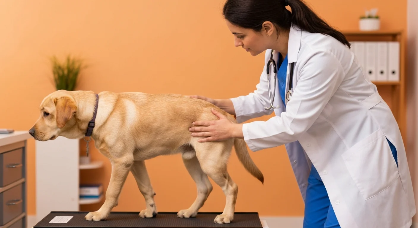
Physical Examination for Early Detection
A systematic elbow examination at every puppy wellness visit from 4 months onward dramatically improves early detection. The examination need not be lengthy, but it must be consistent and specifically targeted at the elbow, which is often overlooked in routine assessments focused on vaccines and deworming.
Systematic Elbow Assessment
- Visual gait analysis: Watch the puppy walk and trot on a level surface before handling. Compare forelimb stride length to hindlimb. Note any asymmetry, circumduction, or head carriage changes.
- Elbow palpation: With the dog standing, palpate both elbows for joint effusion (swelling). Even mild distension of the elbow joint capsule, palpable as a boggy fullness between the lateral epicondyle and olecranon, is abnormal in a young dog and highly suggestive of intra-articular pathology.
- Range of motion: Flex and extend each elbow individually. Note end-range pain (the dog pulls away, turns to look, or tenses), crepitus (a grinding sensation during movement), or reduced range compared to the opposite limb.
- Pronation-supination stress: With the elbow flexed to approximately 90 degrees, rotate the forearm into pronation and supination. Pain on pronation with the elbow flexed is particularly suggestive of medial compartment disease (FCP).
- Comparison: Always examine both elbows, even if lameness appears unilateral. The "sound" limb frequently has subclinical disease that palpation reveals before radiographic changes are detectable.
The Elbow Effusion Test
Joint effusion is one of the earliest and most reliable physical findings in ED. Kirberger and Fourie (1998) demonstrated that palpable elbow effusion in young dogs of susceptible breeds correlates strongly with subsequent ED diagnosis. The test requires practice but becomes straightforward with experience: with the elbow extended, place the thumb and forefinger on either side of the olecranon and lateral epicondyle. A fluid wave or boggy fullness indicates effusion. In a normal young dog's elbow, this area feels firm and bony without soft tissue distension.
When to Pursue Diagnostic Imaging
Physical examination findings determine whether imaging is warranted, but the decision about when and what type of imaging to pursue requires clinical judgment. Not every suspicious finding needs immediate radiographic evaluation, and conversely, some dogs benefit from imaging even when physical findings are equivocal.
Indications for Early Imaging
- Palpable joint effusion in any large breed puppy over 4 months of age
- Reproducible pain on elbow manipulation, particularly on flexion-pronation
- Persistent or progressive lameness lasting more than 2 weeks in an at-risk breed
- Bilateral forelimb stiffness that does not resolve with conservative rest
- Positive family history: Siblings or parents diagnosed with ED significantly increases the index of suspicion
Imaging Considerations
Standard radiographs remain the first-line imaging modality for ED screening. However, early disease may not produce obvious radiographic changes, particularly FCP where fragment visualization requires precise positioning. If clinical suspicion remains high despite normal radiographs, CT scanning provides substantially greater sensitivity for detecting early fragmentation, subchondral bone changes, and joint incongruity. CT has been shown to identify FCP lesions missed by radiography in approximately 30% of cases (Moores et al., 2008).
The timing of imaging matters. Screening before 5 months is rarely productive as the elbow is still developing normally. Between 5 and 8 months, imaging has the highest yield for detecting primary lesions before secondary OA obscures the picture. After 12 months, secondary changes may be the dominant finding, making it difficult to assess primary pathology. The IEWG scoring system is most meaningful when applied during this optimal imaging window.
The Cost of Delayed Diagnosis
Multiple studies confirm that outcomes for surgical intervention are significantly better when ED is diagnosed and treated before advanced secondary osteoarthritis develops. Burton et al. (2011) reported that dogs undergoing arthroscopic fragment removal before 12 months of age had significantly better long-term function scores than those treated after 12 months. Each month of delayed diagnosis during active disease allows progressive cartilage damage that surgery cannot reverse. Early detection is not merely academic; it directly determines whether a dog will walk comfortably for life or develop chronic lameness.
The Role of Breeders in Early Detection
Responsible breeders occupy a unique position in the early detection chain. They see puppies during the critical developmental window, they know the lineage, and they can observe emerging clinical signs before the puppy transitions to its new home.
Breeder Screening Practices
- Puppy evaluation at 6-8 weeks: While ED is not yet detectable at this age, noting puppies with asymmetric limb development, reluctance to bear weight on forelimbs, or abnormal gait provides early data points.
- Post-placement communication: Establishing clear guidance for new owners about what gait abnormalities to watch for and when to contact the breeder or veterinarian. Written puppy care packets should include ED warning signs for at-risk breeds.
- Screening contracts: Incorporating mandatory elbow evaluation at 12-18 months into puppy sale contracts, consistent with breeder certification requirements, ensures that affected dogs are identified and reported back to the breeding program.
- Pedigree tracking: Maintaining databases of offspring elbow scores allows breeders to identify sires and dams that produce higher ED rates, informing future breeding decisions. This retrospective data is often more valuable than screening parents alone, as the polygenic nature of ED means clinically clear parents can still produce affected offspring.
Early Intervention: What Happens After Detection
Early detection only improves outcomes if it leads to timely and appropriate intervention. The management pathway after early ED identification depends on the specific findings and the dog's age.
Mild Clinical Signs, No Radiographic Changes
Dogs showing subtle gait abnormalities or mild palpation findings but no radiographic evidence of ED require monitoring rather than immediate intervention. Reasonable steps include activity modification (avoiding high-impact exercise), nutritional optimization, and re-evaluation in 4-6 weeks. If signs persist or progress, advanced imaging with CT should be considered.
Confirmed Early ED
When imaging confirms ED, the decision between surgical and conservative management depends on the specific pathology, severity, and the presence of mechanical fragments or flaps. In general, early arthroscopic intervention for FCP and OCD provides the best long-term outcomes when fragments or flaps are present. For mild joint incongruity without fragmentation, conservative management with close monitoring may be appropriate.
The Importance of Bilateral Assessment
Because of the high bilateral incidence, any dog diagnosed with ED in one elbow should have the contralateral elbow thoroughly evaluated, even if it appears clinically normal. Subclinical disease in the opposite limb is common and identifying it early allows simultaneous or staged treatment rather than discovering it months later when the "good" leg begins to fail.
Screening Programs and Population-Level Early Detection
Individual case detection is important, but population-level screening programs offer the greatest potential for reducing ED prevalence over time. The screening protocols recommended by organizations like the IEWG, OFA, and BVA/KC are designed to identify affected individuals before they enter breeding programs.
However, current screening recommendations typically specify minimum ages of 12-24 months for formal certification. While appropriate for breeding clearance, this timeline is too late for optimizing individual patient outcomes. A two-tier approach serves both goals: clinical screening at 6-8 months focused on early detection and treatment, followed by formal radiographic certification at the age required by the relevant registry for breeding clearance.
Breed clubs and registries are increasingly recognizing the value of earlier screening. Some progressive programs now encourage preliminary elbow evaluations at 6-9 months, with results flagged for follow-up rather than used for final certification. This approach, mirrored in hip dysplasia screening programs for shepherd breeds, identifies at-risk dogs during the treatment window while preserving the integrity of formal certification at skeletal maturity.
The Economic Case for Early Detection
Early detection is cost-effective even before considering welfare benefits. Early arthroscopic intervention typically costs less than managing chronic osteoarthritis over a dog's lifetime. A single arthroscopy at 8 months may prevent years of NSAID therapy, repeated veterinary visits for lameness management, rehabilitation sessions, and eventual salvage surgery. For breeders, early identification prevents affected dogs from entering breeding programs, reducing the economic and reputational costs of producing clinically affected puppies.
Practical Checklist for Owners of At-Risk Breeds
Owners of large and giant breed puppies can actively participate in early detection by monitoring for the following at each developmental stage:
| Age | What to Monitor | Action if Concerns |
|---|---|---|
| 8-16 weeks | General gait symmetry, willingness to play, weight gain rate | Discuss growth rate with veterinarian; ensure appropriate large breed puppy diet |
| 4-6 months | Morning stiffness, stair reluctance, shortened forelimb stride, splayed sitting posture | Request elbow palpation at next wellness visit; describe specific observations |
| 6-9 months | Exercise intolerance, intermittent lameness, swelling around elbows, head carriage changes at trot | Schedule orthopedic evaluation; radiographs if physical findings present |
| 9-12 months | Persistent stiffness, worsening after exercise, muscle asymmetry in forelimbs | Imaging recommended; consider CT if radiographs inconclusive but suspicion remains |
| 12-18 months | Formal screening per breed club/registry requirements | Submit for official elbow evaluation and certification |
Advances in Early Detection Technology
Research continues to refine early detection methods beyond standard clinical examination and radiography.
- Biomarkers: Synovial fluid and serum markers of cartilage degradation (such as C2C, CPII, and hyaluronic acid levels) show promise for detecting joint pathology before radiographic changes. While not yet in routine clinical use, these markers may eventually allow blood-test screening of at-risk puppies.
- Pressure plate gait analysis: Force plate and pressure mat systems can detect subtle lameness that escapes visual observation. Quantitative gait analysis has been shown to identify limb loading asymmetries in dogs assessed as "sound" by experienced clinicians.
- AI-assisted radiographic interpretation: Machine learning algorithms trained on large databases of elbow radiographs are showing improved sensitivity for detecting early radiographic changes compared to general practitioner interpretation. These tools may be particularly valuable in primary care settings where orthopedic expertise is limited.
- Genetic risk scoring: As genomic research identifies more of the loci contributing to ED, polygenic risk scores may eventually predict individual susceptibility with sufficient accuracy to guide screening intensity. Puppies with high genetic risk scores could be flagged for enhanced monitoring from birth.
Related Database Resources
- Elbow Dysplasia Overview - Complete condition overview
- Screening Protocol - When and how to test
- CT Scanning - Advanced diagnostic imaging
- Breed Prevalence - Which breeds are most at risk
- Rehabilitation - Post-diagnosis physical therapy
Conclusion
Early detection of elbow dysplasia fundamentally changes outcomes. The difference between identifying ED at 6 months versus 18 months can mean the difference between a straightforward arthroscopic procedure with excellent prognosis and managing chronic, progressive osteoarthritis for the remainder of the dog's life. The clinical signs are there to be found, but finding them requires deliberate attention during the developmental period, systematic elbow examination at every puppy visit, and a low threshold for imaging in at-risk breeds. Breeders, owners, and veterinarians each play essential roles in this detection chain. When all three are educated and vigilant, elbow dysplasia can be caught during the narrow window where intervention makes the greatest difference.